Pregnancy Ultrasound

Introduction
A pregnancy ultrasound is used to create images of a developing baby and the pelvic organs of the mother during pregnancy. An ultrasound uses sound waves to create pictures on a video monitor. Ultrasound does not use radiation like X-rays, and there are no known risks associated with the procedure. Ultrasound may be used from the fifth gestational week until delivery. It is an amazing way to view the growth of your developing baby.
An ultrasound uses a small conduction device that is placed on your skin for a pelvic ultrasound or a conduction wand that is inserted into the vagina for a transvaginal ultrasound. The conduction device transmits sound wave information that is translated into pictures on a monitor. Particular images may be saved in the computer or printed out.
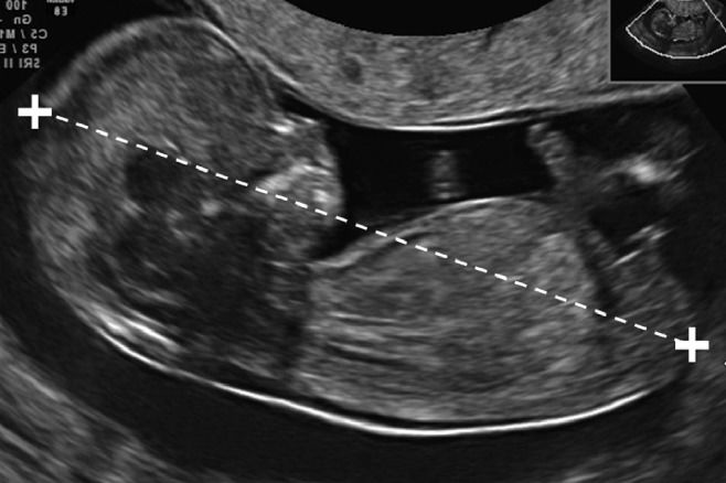
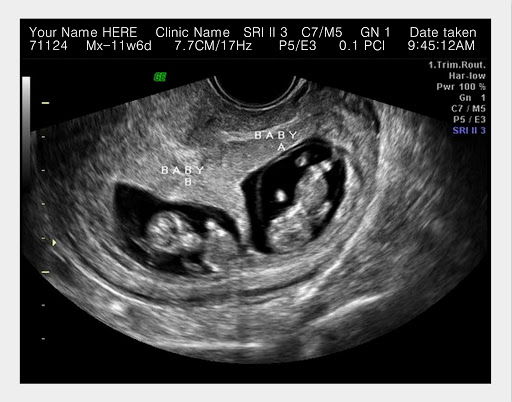
In early pregnancy, an ultrasound may be used to confirm a healthy pregnancy, diagnose multiple pregnancies, and estimate the age of an embryo or fetus. An early ultrasound may be used to check for ectopic pregnancy, sources of bleeding, or signs of miscarriage. At as early as six weeks, a baby’s heart may be seen beating on an ultrasound. In time, an ultrasound may show if a baby is a boy or girl.
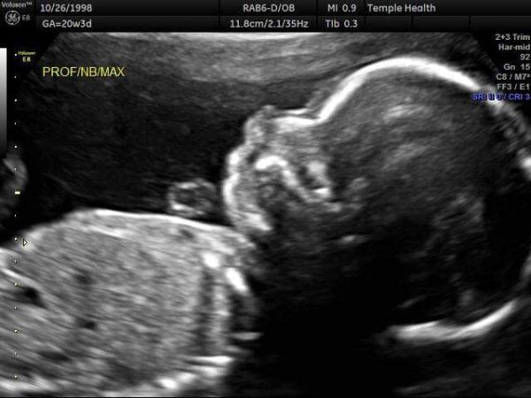
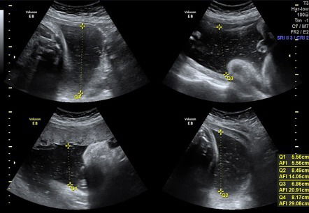
As your baby develops, ultrasounds may be taken to show his or her size and position in your uterus. The condition and location of the placenta and amount of amniotic fluid may be monitored. Ultrasound may be used to check for Down’s Syndrome or other developmental abnormalities. You may be able to see your baby move, breathe, suck his or her thumb, and even hiccup. Prior to delivery, ultrasound can confirm the position of your baby and the umbilical cord to help plan a safe birth.
The amount of ultrasounds that you receive during your pregnancy may depend on several factors. Some doctor’s perform screening ultrasounds, and some perform ultrasounds only when a problem is suspected. Women with high-risk pregnancy may undergo more ultrasounds than those with low-risk pregnancy.
Treatment
An ultrasound is an outpatient examination that may be performed in a doctor’s office, the radiology department of a hospital, or an imaging center. You may be asked to drink several glasses of fluid an hour before your test. A full bladder helps to create a good image for an abdominal ultrasound. You should not urinate before your test unless instructed to do so.
You will lie on your back on an examination table for the procedure. A conducting gel will be placed on your skin. A radiology technician or your doctor will gently place and move the conduction device on your lower abdomen to create the images. An ultrasound usually causes only slight or no discomfort.
A transvaginal ultrasound is performed with an empty bladder. You will undress from the waist down and use a sheet for coverage. You will lie on your back on an examination table and place your feet in stirrups to position your pelvis. The conduction wand is gently inserted into your vagina and positioned to produce the best images. You may feel temporary slight discomfort or pressure during the procedure.
When to expect an ultrasound?
1) 1st trimester- if you have not had a regular period and have a positive pregnancy test, a transvaginal pelvic ultrasound helps to confirm an early pregnancy AND help to correlate the dating criteria to establish your gestational age AND your estimated duet date. This is important to establish when to perform certain tests, when to deliver, etc.
2) Mid trimester- 18-22 weeks of pregnancy anatomy scan, AKA Level 2 scan. This scan is used to confirm appropriate growth, and that the major structures have developed normally. This is also the scan to peek at the baby's sex if not already known from your cell free DNA test. Any abnormalities seen here may prompt a referral to a Maternal Fetal Medicine specialist
3) 36 weeks- this scan may or may NOT be necessary but is done if the position of the baby is not easy to determine by Leopold. it can also look at the thickness of the lower uterine segment in VBAC patients.
Additional scans
-Antepartum testing. Certain women with medical or pregnancy complications warrant scheduled testing to assess fetal well being and growth. This may be required on a weekly basis and start as early at 28 weeks.
-Twin pregnancies These pregnancies require monthly growth scans to ensure both babies are growing appropriately
-Size discrepancy- any signfiicant change in the Symphysis to Fundal height both smaller or greater than expected may prompt a repeat growth scan to see a more accurate representation of the baby's growth.
-Morbidly obese patient may require more frequent sonogram to assess baby's growth
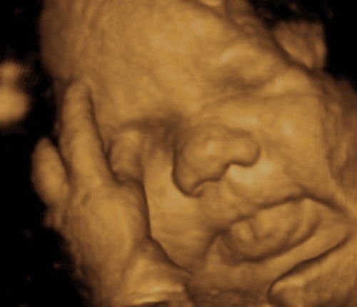
3D scans are not covered by insurers or have been shown to be superior to 2 dimensional scans. These are typically done by patients to get very cool pictures of their baby.
If your doctor performs your ultrasound, your results may be discussed at the time of your test. A radiology technician may perform your test, but is not qualified to diagnose or discuss your results with you. In this case, a radiologist or your doctor will review your images shortly after your test. Your doctor will contact you with the results.